
Ocular Diseases
Many ocular diseases can adversely affect a person's vision. Listed below are some pathologies that can disrupt normal eyesight. Your ophthalmologist will be happy to any questions that you have about your condition.

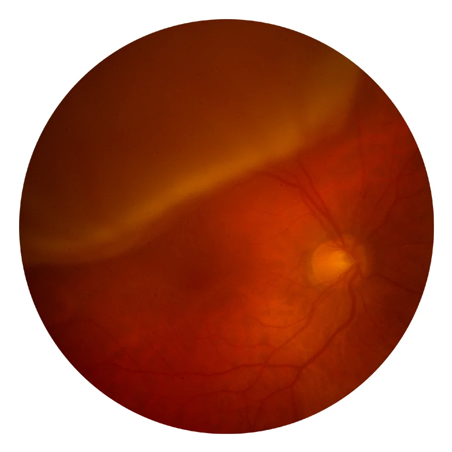
Diabetic Retinopathy
Central Serous Retinopathy
Central Serous Retinopathy (CSR) is a pathological condition where a blister-like elevation occurs in the center area of vision (macula). Small breaks in the cell layer outside the retina allows fluid to leak into the macula resulting in distorted or blurred vision.
A fluorescein angiogram (dye test) may be used to confirm the diagnosis. In some cases, a smokestack shape will
appear. While the cause is unknown, most patients regain vision within several months.
Cystoid Macular Edema
Cystoid macular edema (CME) is a pathological condition where cysts form in the area of central vision causing retinal inflammation. Distorted or blurred vision is a common symptom of CME, while the periphery (side vision) is not affected by this disorder. The decreased vision associated with CME is usually temporary; however, it can recur.
The exact causes for CME are unknown, but it frequently occurs following cataract surgery. Treatment options may include anti-inflammatory drugs (such as steroids), laser or vitrectomy surgery.
Diabetic Retinopathy
Diabetic Retinopathy is a pathological condition which produces a series of steadily advancing retinal changes in patients with diabetes mellitus. Early stages of retinopathy may produce leaking blood vessels and swelling of the retina. Advanced stages may include the growth of abnormal blood vessels, which may damage the eye and lead to glaucoma, retinal detachment or other sight-threatening conditions.
Patients may experience blurred vision or have no symptoms whatsoever as the disease progresses. Treatment options include laser to reduce swelling and shrink the abnormal vessels.
Drusen
Drusen are tiny glassy particles that appear behind the retina on the innermost part of the choroid (the layer of the retina containing blood vessels). This anatomic defect commonly occurs in patients over the age of 60.
Drusen sometimes presents as an early sign of the onset of age-related macular degeneration.
Retinal Detachment
Geographic Atrophy
Geographic atrophy (GA) is the advanced stage of "dry" age-related macular degeneration. "Geographic" describes the irregular-shaped areas where loss of pigment has occurred. Atrophy refers to the wasting away of the cells, in this case, in the retinal pigment epithelium (RPE) layer which nourishes visual cells.
This condition has a tendency to progress slowly, and is more common in Caucasians, cigarette smokers, and in individuals with a light-colored iris.
Macular Hole
A Macular Hole is a pathological condition where an abnormal opening occurs in the center of vision. The most common cause of a macular hole is pulling or tugging on the retina by the vitreous, which is the gel-like substance that fills the inner eye. Sometimes scar tissue on top of the retina could also cause the retina to tear.
Symptoms may include blurred or distorted vision, or patients may also experience a blind spot.
Macular Pucker
(Also known as: epiretinal membrane, cellophane maculopathy or epimacular proliferation)
A macular pucker is a pathological condition that distorts the vision. The transparent membrane lying on the retinal surface becomes wrinkled in the macular area due to contraction of the membrane.
Melanoma
A melanoma is a malignant (life-threatening) dark-pigmented tumor. Although rare, they can grow in nearly all the tissues within the eye.
Treatment options include observation, radiation treatment, laser treatment, and enucleation (removal of the eye). Even with treatment a melanoma may spread (metastasize) to other parts of the body.
Retinal Detachment
Retinal Detachment is a pathologic condition in which the retina is separated from the underlying cell layer.(back wall of the eye). It most often is caused when a tear occurs in the retinal tissue and fluid in the eye leaks behind the tear.
Symptoms may include loss of peripheral vision, flashes of light or floaters (moving particles that cast shadows on the retina). Retinal detachment almost always needs immediate surgical treatment.

Branch Retinal Vein Occlusion
Retinal Tear
Tears can form in the retinal tissue. Often as we age, the gel-like substance in the inner eye (vitreous) shrinks and can tug at the retina causing it to tear. A tear can lead to a retinal detachment in which the retina separates from the back of the eye, causing a severe loss of vision.
While there is no way to predict if a retinal tear will occur, some other risk factors include trauma, previous eye surgery, and a family history of tears or detachment.
Retinal Vein Occlusions
A Branch Retinal Vein Occlusion (BRVO) is a pathological condition where blockage of blood flow occurs in a tributary of the central retinal vein. Usually it will cause a decrease in vision. Often the blockage is caused by an hardened, overlying artery. With BRVO, flame-shaped hemorrhages, fluffy, white patches (cotton wool spots), macular edema (swelling) and retinal vein engorgement may be present.
A Central Retinal Vein Occlusion (CRVO) is a pathological condition where a blockage of blood flow occurs in the central retinal vein and causes a decrease in vision. With CRVO, retinal exams may show: retinal thickening, enlarged veins, retinal hemorrhages and edema in the optic nerve margins. Vision may slowly improve over time.
Retinitis Pigmentosa
This pathologic condition is a hereditary disease that causes progressive retinal deterioration in both eyes. The most common symptoms of retinitis pigmentosa are night blindness and loss of peripheral (side) vision.
Presently, there is no specific treatment for retinitis pigmentosa. Patients with this condition experience progressive visual loss and can develop other diseases, such as glaucoma and cataracts.
Vitreous Hemorrhage
A vitreous hemorrhage is a pathological condition where blood leaks into the central cavity of the eye. It may be caused by the formation of new, abnormal blood vessels (neovascularization), trauma, vitreous detachment or retinal tear.
Additionally, hemorrhages or bleeding may be found in other layers and areas of the eye. Hemorrhaging can be a symptom of other existing conditions.